Tag: x-rays

Online Consultations – Which X-Rays Do I Need?

X-rays are critical for diagnosing and managing Scoliosis. They are used to diagnose scoliosis by taking an image of the spine from different angles, which allows specialists to measure the degree of curvature, identify the location and type of scoliosis, and track the progression of the condition over time. For scoliosis to be diagnosed, there must be a measurable curve in the spine which is over 10 degree’s.

X-rays are also used to monitor the effectiveness of scoliosis treatment, during bracing or exercised based therapy, X-rays are used to monitor the response of the curve to treatment, and adapt the treatment plan to ensure that the spine is responding as expected.

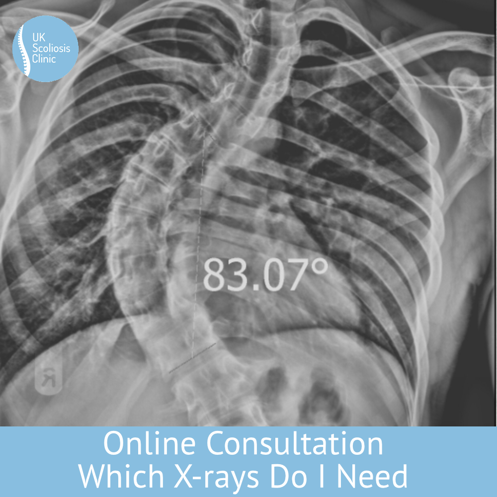
Which X-Rays do I need?

If you’re planning on booking an online consultation at the UK Scoliosis Clinic, it can be very helpful to have an X-ray to send us. You don’t have to have one – but as you can imagine we can give you better information if you do have one to hand. While those who attend the clinic in person can have X-rays taken here, those attending remotely will often need to arrange their own X-rays, or at least request copies from their medical doctor or hospital. So which ones are most useful from a Scoliosis point of view?

The correct type of X-ray to request for a Scoliosis consultation is a standing full spine X-ray taken at 180cm focal distance. Typically, two X-ray images are needed: one from the back (posterior-anterior or PA view) and one from the side (lateral view). It is also important to make sure that the pelvis and hips are in the images. Laying on your back x-rays are not accurate for scoliosis diagnosis and measurement, as they do not allow for the effect of gravity on the curve when standing.

The two X-ray images provide healthcare professionals with crucial information to measure the degree of curvature of the spine. The Cobb angle is the most commonly used method for measuring the degree of curvature and is calculated by adding the slant/slope of the most tilted vertebrae at the top and most tilted vertebrae at the bottom of the curve together. A Cobb angle of 10 degrees or less is considered within the normal range, whereas a Cobb angle of 10-25 degrees is classified as mild scoliosis, 25-40 degrees is moderate scoliosis, and 40 degrees or more is severe scoliosis.

As you can imagine, having the ability to determine the Cobb angle is a critical aspect of a Scoliosis diagnosis, and this is why having an X-ray to have for your consultation allows us to provide you with much more certainty.  To confirm the diagnosis of structural scoliosis we need to confirm a Cobb angle of 10 degrees or more, combined with vertebral rotation. Without X-rays, we can still give a professional opinion – however, especially for smaller curves an X-ray is often the most important item to have so far as diagnosis is concerned.


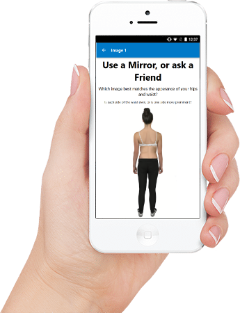
If you don’t have X-Rays

If you don’t have X-rays you’re still more than welcome to book an online consultation – there’s still a great deal of advice we can give even without X-rays, however, if you can get your hands on an X-ray it’s a huge advantage.

If this is an issue for you, we would recommend that you consider an in-clinic consultation instead, if possible. At the clinic, your appointment can include a full set of Diagnostic X-rays utilising our state-of-the-art digital X-ray machine, allowing Scoliosis to be definitely diagnosed and understood. You will also receive a digital copy of your X-rays to take away.

Our in-clinic consultations are generally the most suitable for those who are concerned that they may have scoliosis but do not have X-rays or other documentation to rule the condition in, or out. It’s also ideal for those who know they have Scoliosis and are actively looking to take up non-surgical treatment or are wanting to change treatment, perhaps from another provider.


Should I remove my brace before a progress X-ray?

X-rays are the gold standard when it comes to diagnosing scoliosis and tracking the progress of treatment for the condition. Scoliosis is notoriously difficult to gauge from a visual inspection alone, meaning that regular X-rays are critical to ensuring that treatment is progressing as expected and that any adjustments to a patient’s brace are made at the correct time. In the usual course of treatment, Scoliosis professionals take X-rays every 6-12 months with these goals in mind – which often leaves patients asking, should I take my brace off before the X-ray?


In-brace vs out of Brace correction

What’s important to understand is that a Scoliosis Brace provides better correction while being worn than not being worn – that might sound obvious but it’s important to remember that the role of the brace is to correct, or often, over correct Scoliosis, in order that over time the spine is corrected when the brace is removed.

When you put a Scoliosis brace on, research suggests that it takes around 2 hours[1] to reach the maximum correction that can be obtained by using a brace. After removing, the correction obtained is gradually lost over around 2 hours, after which the curve/spine stabilizes[2]  Over time, the goal of Scoliosis treatment is to reduce the amount that correction is lost when the brace is removed – over corrective braces, such as ScoliBrace achieve this by adding corrective force to he spine to help to move it towards its proper position.


Braces and X-rays

So, should you remove your brace before a progress X-ray?  Taking the brace off, perhaps even days before the X-ray gives a truer picture of what’s “really” happening with the spine – but tells us little about how much correction the brace itself is providing. By contrast, leaving the brace on during the X-ray, or removing it right before gives us an excellent idea of how well the brace is working (and whether it’s time to adjust it), but isn’t as good at showing us how the spine might look after hours, or days of no wear. A good compromise is often an in-brace X-ray, followed by an out-of-brace X-ray after a short break which can give an idea of in-brace correction, and allow us to approximate an out-of-brace figure.

Nonetheless, it’s a complex issue, and for these reasons, at present, there’s no defined standard for the best way to carry out progress X-rays – generally speaking, it’s up to the Scoliosis practitioner to decide how to perform X-rays, based on the specific needs of the patient.

But let’s not forget the most important fact about Scoliosis bracing – it’s a treatment method which works over time – for this reason, we’re often less concerned with the exact method used to take progress X-rays, and more with the fact that the x-rays are performed in a consistent way. That is to say, during treatment, we care less about the exact degree of correction and more about the direction of travel.


Should I remove my brace before a progress X-ray?

The answer to the question is simply… do as your practitioner suggests! When to remove, or not remove the brace isn’t an issue that you as a patient need to worry about – but you should make sure that you comply with the instructions you’re given, and that you do so each time you visit for a progress X-ray. Rember, it’s consistency which matters!


[1] Katarzyna Zaborowska-Sapeta et al. The Duration of the correction loss after removing cheneau brace in patients with adolescent idiopathic scoliosis Acta Orthopaedica et Traumatologica Turcica 53 (2019)

[2] Meng Li  1 , M S Wong, Keith D K Luk, Kenneth W H Wong, Kenneth M C Cheung, Time-dependent response of scoliotic curvature to orthotic intervention: when should a radiograph be obtained after putting on or taking off a spinal orthosis?  Spine (Phila Pa 1976). 2014 Aug 1;39(17):1408-16.